Preliminary Phytochemical Screening, Isolation, Characterization, Structural Elucidation and Antibacterial Activities of Leaves Extracts Rhus vulgaris (Kimmo)
Aderaw Anteneh Belew1*  , Getachew Mariam Hana2 and Desta Shumuye Meshesha2
1Department of Chemistry, College of Natural and Computational Sciences, Jigjiga University, Jigjiga, Ethiopia .
2Department of Chemistry, College of Natural and Computational Sciences, University of Gondar, Gondar, Ethiopia .
http://dx.doi.org/10.13005/OJPS09.02.08
Rhus vulgaris, commonly known as sumac, is a plant known for its various therapeutic properties, including antioxidant and antibacterial activities. Medicines derived from plants significantly contribute to human health. This study aimed to screen the phytochemical constituents, isolate and elucidate the structure, and evaluate the antibacterial activity of the methanol extract from the leaves of Rhus vulgaris. The concentrated fraction was purified using thin-layer chromatography (TLC) and column chromatography to isolate the pure compounds. The isolated compounds were characterized using Fourier transform infrared spectroscopy (FTIR) and nuclear magnetic resonance spectroscopy (NMR). Antibacterial activity was tested against four bacterial strains: Streptococcus aureus (gram-positive), Escherichia coli, Salmonella typhimurium, and Klebsiella pneumoniae (gram-negative) using the agar well diffusion method. The methanol extract exhibited antibacterial activity against all tested bacteria, with significant inhibition zones, particularly against Streptococcus aureus (15 mm) and Salmonella typhimurium (14 mm). The preliminary phytochemical screening of the extracts revealed the presence of alkaloids, glycosides, steroids, anthraquinones, and carbohydrates were detected in all extracts. The methanolic extract of Rhus vulgaris was subjected to column chromatography and eluted with methanol: chloroform (1:8) mixture, leading to the identification of the compound 1-p-tolyl pentadeca-7,9-dien-1-ol. The methanolic extract of Rhus vulgaris has demonstrated strong antibacterial activity, indicating its potential as an effective antimicrobial agent.
Copy the following to cite this article:
Belew A. A, Hana G. M, Meshesha D. S. Preliminary Phytochemical Screening, Isolation, Characterization, Structural Elucidation and Antibacterial Activities of Leaves Extracts Rhus vulgaris (Kimmo). Oriental Jornal of Physical Sciences 2024; 9(2).
DOI:http://dx.doi.org/10.13005/OJPS09.02.08Copy the following to cite this URL:
Belew A. A, Hana G. M, Meshesha D. S. Preliminary Phytochemical Screening, Isolation, Characterization, Structural Elucidation and Antibacterial Activities of Leaves Extracts Rhus vulgaris (Kimmo). Oriental Jornal of Physical Sciences 2024; 9(2). Available here:https://bit.ly/4f7QHIh
Download article (pdf) Citation Manager Publish History


Introduction
Natural products, which contribute nearly 50% of new chemical entities in drug development, are crucial in providing starting points for synthetic drugs1. Medicinal plants have been used since ancient times for various purposes and continue to guide the search for new medications due to their accessibility, affordability, and minimal side effects compared to synthetic drugs. These plants contain phytochemicals that produce specific physiological actions2. The medicinal efficacy of plants is attributed to these chemical substances, known as phytochemicals, which exert definite physiological effects on the human body3,4.
The Rhus genus, known as sumac, comprises over 250 flowering plant species in the Anacardiaceae family5, predominantly found in tropical, subtropical, and temperate regions worldwide. The name "sumac" originated from the Syriac word "sumaga," meaning red. These non-agricultural plants have been utilized by indigenous people for medicinal and other purposes, indicating their potential for commercial application without competing with food production6. Traditional Rhus species have been employed to treat various ailments, including influenza, wounds, diarrhea, stomach pain, indigestion, diabetes, malaria, rheumatism, gum and toothaches, swollen legs, dog bites, peptic ulcers, kidney stones, skin eruptions, bruises, and boils7,8. In Ethiopia, R. vulgaris is used to manage various diseases according to ethnobotanical and ethno-pharmacological research, such as diarrhea, gonorrhea, infections, the evil eye, wounds, and lung tuberculosis in the Amhara region 9,10. In Kenya, this plant’s stem bark is used to treat malaria11. In Tanzania, Fresh leaves are burned and the ash is used for oxytocic action, and externally, the ash is applied for the treatment of scabies. The plant is used to stop diarrhea, wounds, gonorrhea, and infertility, and to ease delivery12. In Uganda, this plant is used to treat diarrhea, malaria, hemorrhoids13, Yellow fever, Cough, Malaria, Gastro-intestinal disorders, toothaches, Syphilis, Immunity booster, Smallpox, Swollen lymph, and Prevention diseases14, more preferable chewing sticks over synthetic toothbrushes 15. In Kenya, the roots, leaves, and fruits are used as the treatment of cancers like Stomach, skin, and breast cancer 16.
In recent studies, researchers have successfully isolated and characterized novel compounds from Rhus species, demonstrating their therapeutic potential. These findings underscore the importance of further exploring Rhus vulgaris for its pharmacological benefits and developing these natural products into clinically valuable drugs. Phytochemical studies on Rhus vulgaris stem bark have identified tannins, saponins, flavonoids, terpenoids, glycosides, alkaloids, and phenols. The methanolic extract showed superior anti-inflammatory activity compared to indomethacin17. Other studies have identified new biflavonoids in Rhus species, including agathisflavone, amentoflavone, hinokiflavone, rhus flavanone, and succedanea flavone, which exhibit activity against various significant viruses. Hinokiflavone emerged as the most active among 65 natural flavonoids in inhibiting the pro-coagulant activity of adherent human monocytes stimulated by endotoxin and interleukin-1-? in vitro. Additionally, other Rhus biflavonoids demonstrate cytotoxic and antimalarial activities 18,19.
This study aims to isolate potentially bioactive compounds from Rhus vulgaris leaves using column chromatography and preparative thin-layer chromatography. Structural elucidation of the active compound will be conducted using spectroscopic techniques such as FTIR and NMR. Furthermore, the study seeks to assess the bioactivity of Rhus vulgaris leaves against various pathogens.
Description
Rhus vulgaris is found in all parts of Tanzania; Uganda and Kenya and from Cameroon to Ethiopia and south to Mozambique, Malawi, Zambia, and Zimbabwe20. It is a shrub or small tree that occasionally reaches 1-9 m; its bark is smooth, dark brown, its branches yellow-red-brown, and often densely hairy. Leaves are 3 leaflets, dull green, softly hairy, the central leaflet larger, 4.11 cm long x 2.6.5 cm wide, the two laterals smaller, shortly stalked, edge entire or soft toothed towards the tip, which is blunt or pointed, leaflets dark above, paler below. Flowers are small cream-green-yellow, parts in fives, in terminal loose heads or from upper leaf axils, 5.20 cm long, all densely hairy. Fruits are drupes, with thin flesh, flat and round, red-brown, and only 3.5 mm across21.
Taxonomic Classification
According to the International Code of Botanical Nomenclature, the present taxonomic classification of Rhus vulgaris is shown in Figure 1:
Kingdom Plantae
Subkingdom Tracheobionta
Superdivision Spermatophyta
Division Magnoliophyta
Class Magnoliopsida
Subclass Rosidae
Order Sapindales
Family Anacardiaceae
Genus Rhus
Species Rhus vulgaris Meikle
Synonyms
Searsia pyroide (Burch.) Moffett
Rhus pyroide (Burch.) 10
Materials and Methods
Reagents and Chemicals
All the chemicals and reagents used in the present study were of analytical grade. The chemicals and reagents included: Methanol (98%), acetic anhydride, ferric chloride solution, ammonia, Mayer’s reagent, Benedict’s reagent, Sodium bicarbonate, ammonia, Ninhydrin, Molisch’s reagent, Millon’s reagent, lead acetate, Folin-Ciocalteu reagent, gallic acid, ascorbic acid, sodium hydroxide, anhydrous sodium carbonate, and anhydrous aluminum chloride, which were purchased from Loba Chemie (Mumbai, India); whereas sulfuric acid, acetic acid, ethanol, n-hexane, chloroform, acetone, ethyl acetate, DMSO, Vanillin solution (15g of vanillin in 250 ml of ethanol and add 2.5 ml of conc. H2SO4), NaOH, concentrated hydrochloric acid, silica gel (60-200 Mesh size) from (Oxford Lab. Chem., India), distilled water, are more relevant to make this experiment.
Apparatus and Instruments
The apparatus and instruments that were used for the study include: electrical grinder (IAK–WERKE, Germany), electronic balance (Bosch, Germany), Whatman No. 42 filter paper (110 mm), conical flasks, volumetric flasks, TLC plates, measuring cylinders, graduated pipettes, micropipettes (Mumbai, India), refrigerator (Hitachi LR902T, USA), orbital shaker (GEMMY Orbital Shaker, VRN-480, Taiwan), Autoclave, balance (Sartorius, Germany), rotary evaporator (Bibby RE200, Sterilin Ltd., UK) and UV/Vis spectrophotometer (Sanyo SP75, UK) were used in the study.
FTIR: The isolate was mixed with 200 mg KBr (FT-IR grade) and pressed into a pellet. The sample pellet was placed into the sample holder and FT-IR spectra were recorded in the range 400- 4000 cm-1 in FT-IR spectroscopy (Bruker FTIR Spectrometer, USA).
Nuclear Magnetic Resonance Spectroscopy
The 1H, 13C-NMR, and 1D NMR spectra of base degradation impurities were recorded in DMSO-d6 solvent on Bruker 400 MHz Avance -III HD NMR spectrometer equipped with broadband observe (BBO) probe. The 1H and 13C chemical shifts are reported on the ? scale in ppm, relative to tetramethyl silane (TMS) as an internal standard. The spectra were set to ? 0.00 ppm in 1H NMR (TMS) and ? 39.50 ppm in 13C NMR (DMSO-d6).
Collection of plants
The fresh and healthy leaves of R. vulgaris were collected from a local farm located in the Region of Amara, specifically in Takusa Woreda Kebele 12, Central Gondar Zone, in February 2020. Taxonomic identification of the plant was conducted by botanist Mr. Getenet Chekol (MSc.) at the Department of Biology, University of Gondar.
After collection, the plant materials underwent thorough washing with tap water to eliminate any dust particles adhering to the leaves. Subsequently, the collected plant material was air-dried in the shade and stored at room temperature for 10 days. The dried leaves, which were in good condition, were then ground into a uniform powder using an electric grinder. The powdered samples were accurately weighed and stored in sealed containers for subsequent extraction purposes.
 | Figure 1: The photograph of Rhus vulgaris Leaves. |
Extraction
The organic compounds present in Rhus vulgaris leaves were extracted and isolated using the maceration method, chosen for its ability to thoroughly extract the leaves without risking the thermal decomposition of any heat-sensitive compounds22. The powdered leaves (800g) underwent sequential maceration with solvents of increasing polarity: n-hexane (C6H14) 2L, chloroform (CHCl3) 2L, and methanol (CH3OH) 2 L, each for approximately 72 hours. Regular shaking ensured complete extraction as shown in Figure 2. The resulting extracts were decanted, filtered with Whatman filter paper, and concentrated using a rotary evaporator at temperatures specific to each solvent (n-hexane: 69?, chloroform: 61?, methanol: 65?). The crude extracts were then weighed using a digital balance, yielding n-hexane (4.35 g), chloroform (14.7 g), and methanol (100.7g) extracts. Among these, the methanol extract exhibited superior antibacterial activity and underwent further purification via column chromatography, using a mixture of methanol: chloroform (1:8) determined through TLC trials to isolate the major component, as detailed in Table 1.
Column Chromatographic
The methanol extract underwent silica gel (60-120 mesh ASTM, Merck) glass column chromatography with a diameter of 20-25 mm. Initially, silica gel (150 g) was mixed with chloroform to form a homogeneous slurry, which was then poured into the glass column after stirring to remove bubbles. For sample loading, 6g of the extract was dissolved in 40 ml of methanol. To this solution, 10 g of silica was added and mixed thoroughly by stirring with a glass rod, followed by drying at room temperature. The resulting dried silica-extract mixture was layered onto the column bed.
The column was first eluted with a mobile phase consisting of methanol: chloroform in a 1:8 ratio and allowed to run until a consistent flow was achieved. A total of 59 fractions were collected during elution in Table 1. The profiles of column fractions were monitored by TLC to ensure similarity based on the number and appearance of spots on the plate, visualized using a UV lamp and vanillin solution.
Subsequently, the eluted compound was crystallized and subjected to Nuclear Magnetic Resonance (NMR) analysis (1H-NMR, 13C-NMR, DEPT-135) and Fourier transform infrared (FTIR) spectroscopy to elucidate its structure. The compound isolated from this fraction of column eluate was observed to be dark green and gummy in appearance.
Table 1: Solvent system used to separate AA1.
No- | Solvent | Solvent Ratio | No. fractions | Code of Fraction | Rf value |
1 | MeOH: CHCl3 | 1:8 | 1-3 | A1 | 0.9 |
2 | MeOH: CHCl3 | 1:8 | 4-16 | A2 | 0.85 |
3 | MeOH: CHCl3 | 1:8 | 17-45 | A3 | 0.70 |
4 | MeOH: CHCl3 | 1:8 | 46-56 | A4 | 0.75 |
5 | MeOH: CHCl3 | 1:8 | 57-59 | A5 | 0.8 |
Preliminary Phytochemical Screening
Phytochemical screening was conducted to evaluate the qualitative chemical composition of crude extracts, employing standard precipitation and coloration methods. This screening aimed to identify major natural chemical groups such as alkaloids, phenolic compounds, glycosides, carbohydrates, flavonoids, saponins, terpenoids, anthraquinones, tannins, steroids, amino acids, coumarins, and proteins. Through these analyses, the presence or absence of these compounds in the tested crude extracts was determined. The crude extracts, previously prepared and stored in a refrigerator, were utilized for these phytochemical tests 23–27.
Antibacterial Activities of the Leaves extracts of Rhus vulgaris
To assess the antimicrobial activity of the crude extract, the following test organisms were employed: Streptococcus aureus (gram-positive), Escherichia coli, Salmonella typhimurium, and Klebsiella pneumoniae (gram-negative) bacteria. The antibacterial activity of both crude and purified extracts was determined using an agar well diffusion assay.
Mueller Hinton Agar (MHA) was utilized as the growth medium. Preparation involved dissolving 19 g of media powder in 500 ml of distilled water, followed by autoclaving at 121°C for 15 minutes to ensure sterilization. The cooled media was poured into plates, allowed to solidify, and positioned upright in the incubator to prevent contamination.
The bacterial cultures were grown on MHA, and inoculums were prepared for the antibacterial assay. Fresh cultures were picked from overnight growth, suspended in 3-4 ml of physiological saline in sterile test tubes, and adjusted for turbidity to match a 0.5 McFarland standard, corresponding to a bacterial load of approximately 1x10⁸ CFU.
Antibacterial activities of Rhus vulgaris extracts were evaluated against the four bacterial strains using the agar well diffusion method. The test bacteria were swabbed onto the surface of the leveled media and allowed to dry for 10 minutes. Wells were then bored into the plates using a sterilized well borer (6 mm diameter), and 100 mg/ml of each extract (n-hexane, chloroform, and methanol) dissolved in DMSO was added. Gentamicin discs (30 mcg/disc) served as the positive antibiotic control. Petri dishes were incubated at 37°C for 24 hours. After incubation, the zones of inhibition were measured and the average inhibition zone diameter was recorded in millimeters.
 | Figure 2: Method of extraction of the plant material. |
Results and Discussion
The yield of solvent extract and isolation of Leaves of Rhus vulgaris
The dried and powdered roots (800 g) of Rhus vulgaris underwent exhaustive extraction, successively employing n-hexane, chloroform, and methanol. The solvent from each extract was recovered under reduced pressure using a rotary evaporator, resulting in n-hexane (4.35 g), chloroform (14.7 g), and methanol (100.7 g) extracts as shown in Table 2. Chromatographic purification of the methanol extract (7 g) yielded a compound coded as AA1. The structure of this compound has been elucidated based on spectroscopic evidence, as described in the following section.
Table 2: Percentage Yields of the Extracts.
Extracts | Weight | Yield (%) |
n-hexane | 4.35g | 0.54 |
Chloroform | 14.7g | 1.84 |
Methanol | 100.7g | 12.6 |
Phytochemical screening of the Leaves Extracts of Rhus vulgaris
Phytochemical analysis of each crude extract from Rhus vulgaris indicated the presence of various pharmacologically significant secondary metabolite classes. Alkaloids, glycosides, steroids, anthraquinones, and carbohydrates were detected in all extracts. Phenols, flavonoids, tannins, coumarins, and proteins were exclusively found in methanol extracts, while terpenoids, saponins, and amino acids were absent in all extracts, as summarized in Table 3.
Table 3: Phytochemical constituents of the leaves extract of Rhus vulgaris.
Phytochemical constituent | n-hexane | Chloroform | Methanol |
Alkaloids | + | + | ++ |
Phenols | - | - | ++ |
Glycosides | + | + | ++ |
Flavonoids | - | - | ++ |
Terpenoids | - | - | - |
Steroids | + | + | + |
Carbohydrates | + | + | + |
Anthraquinones | + | + | + |
Proteins | - | - | + |
Coumarins | - | - | + |
Tannins | - | - | + |
Saponins | - | - | - |
Amino acid | - | - | - |
Key: (+) = Present, (-) = Absent
Antibacterial Activity of the Crude Extracts against Bacterial Strain
The antibacterial activity of Rhus vulgaris extracts using different solvents was evaluated against selected pathogens (Table 4). The methanol extract exhibited robust activity, showing inhibition zones of 15 mm against Staphylococcus aureus, 14 mm against Salmonella typhi, 11 mm against Klebsiella pneumoniae, and 13 mm against Escherichia coli. In contrast, n-hexane and chloroform extracts showed limited effectiveness, with n-hexane displaying 3 mm against S. aureus and chloroform exhibiting 7 mm against both S. aureus and E. coli in Figure 3 and Figure 4.
These results underscore the potent antibacterial potential of the methanol extract from Rhus vulgaris leaves, particularly effective against S. aureus. However, S. typhi and K. pneumoniae exhibited partial resistance to the extracts.
Table 4: Antibacterial efficacy of extracts against pathogens
Extract | Inhibition zone(mm) against | |||
S. aureus | S. typhi | K. pneum | E. coli | |
n-hexane | 3 | 0 | 0 | 2 |
CHCl3 | 7 | 0 | 0 | 7 |
MeOH | 15 | 14 | 11 | 13 |
Ge | 18 | 17 | 14 | 15 |
DMSO | 0 | 0 | 0 | 0 |
The methanol extract's superior antibacterial properties align with previous research, which has consistently demonstrated the potent antimicrobial potential of methanol as a solvent for phytochemical extraction. This efficacy is attributed to methanol's ability to solubilize a broad spectrum of bioactive compounds, thereby enhancing the extract's antimicrobial action 17,28.
The study's outcomes corroborate the traditional use of Rhus vulgaris in treating infections and highlight its potential as a source of natural antibacterial agents. The extracts were more effective against gram-positive bacteria, a trend observed in other studies examining the antibacterial properties of plant extracts29,30. The methanol extract's effectiveness suggests it contains a higher concentration of active phytochemicals capable of penetrating the peptidoglycan layer of gram-positive bacteria29,31.
 | Figure 3: Zone of inhibition of MeOH compared with an inhibition zone of standard antibiotic Gentamicin on S.aureus, S.typhi, K. pneumonia, and E.coli. |
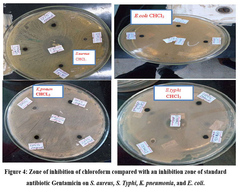 | Figure 4: Zone of inhibition of chloroform compared with an inhibition zone of standard antibiotic Gentamicin on S. aureus, S. Typhi, K. pneumonia, and E. coli. |
Characterization of Compounds
In characterizing the compounds isolated from the leaves of Rhus vulgaris, we utilized both the RF value and spectroscopic data of the compounds.
Partial Characterization of AA1
Compound AA1, a dark green gummy substance, was isolated from the leaves of Rhus vulgaris. It exhibited a pink hue under a UV lamp and turned yellow upon exposure to 4% vanillin H2SO4. In methanol: chloroform (1:8), its Rf value was measured at 0.70.
FTIR Spectral Analysis
The FTIR spectrum of compound AA1 (Figure 5) revealed an absorption band at 3454 cm-1, indicating the presence of the -OH group. Additionally, bands observed at 3000 cm-1, 2912 cm-1, and 2882 cm-1 corresponded to the stretching of aliphatic =C-H, CH2, and CH3 groups, respectively. Furthermore, a band at 1637 cm-1 indicated C=C bending. The bands observed at 1464 cm-1, 1355 cm-1, and 1078 cm-1 were assigned to CH2 bending, CH3 bending, and C-O stretching vibrations, respectively.
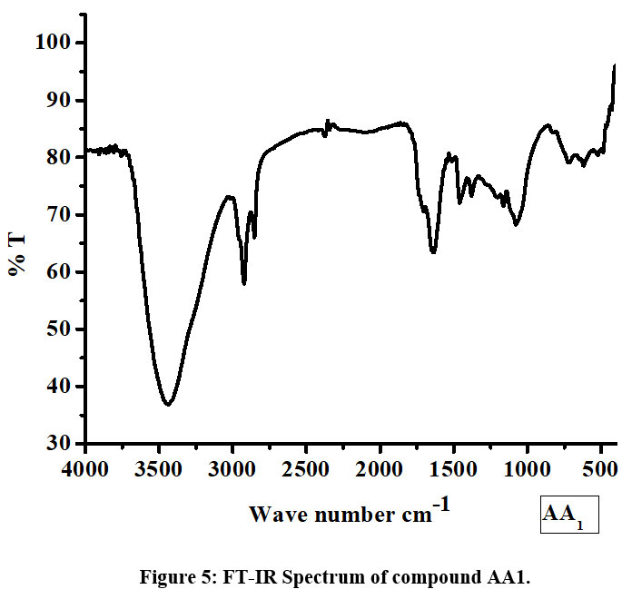 | Figure 5: FT-IR Spectrum of compound AA1. |
1H-NMR Spectrum
In Figure 6, the peak at ? 1.25-2.07 (m) corresponds to the protons of the nine methylene groups, appearing as a multiplet due to signal overlap. Additionally, peaks at ? 0.84 (t) and ? 2.35 (d) indicate the signals of the methyl group protons. The peak at ? 4.50-6.03 (t) indicates the protons of the five methine groups, while ? 6.99-7.07 (d) indicates the protons of the four methine groups.
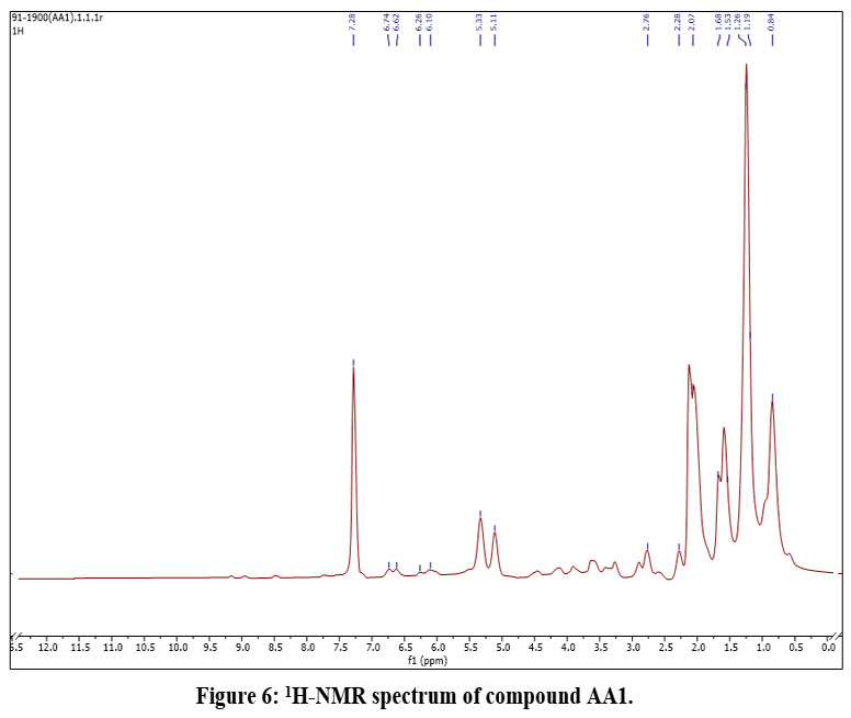 | Figure 6: 1H-NMR spectrum of compound AA1. |
13C-NMR Spectrum
The proton-decoupled 13C-NMR spectrum in Table 5 and Figure 7 displayed signals for 17 carbon atoms, whereas the proposed structure (AA1) contains 22 carbon atoms. The variance in the number of carbon atoms between the proposed structure and the signals detected in the 13C NMR spectrum might be attributed to the presence of chemically or magnetically equivalent carbon atoms. As depicted in Figure 9, carbon atoms 4, 5, 12, 6, 11, 8, 9, 10, 3', 1', 4', and 2' & 6' could be chemically equivalent.
The DEPT spectrum in Figure 8 exhibited signals for 11 carbon atoms, with 4 signals indicating the presence of 9 methylene groups and the remaining 7 signals for CH and CH3 groups. In the DEPT spectrum, data are acquired in a manner that results in signals being either upfield (CH & CH3) or downfield (CH2), depending on the number of attached protons. In contrast, the proton-decoupled 13C NMR spectrum depicted in Figure 7 displayed signals for 22 carbon atoms, while the DEPT-135 spectrum revealed an overlap of signals for 20 carbon atoms.
The disparity in signals between the two spectra suggested the presence of 2 quaternary carbon atoms that are typically not observed in the DEPT-135 spectrum. The 13C and DEPT chemical shifts of the proposed structure are summarized in Table 5 below.
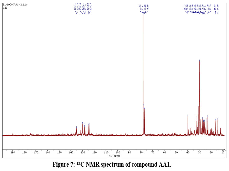 | Figure 7: 13C NMR spectrum of compound AA1. |
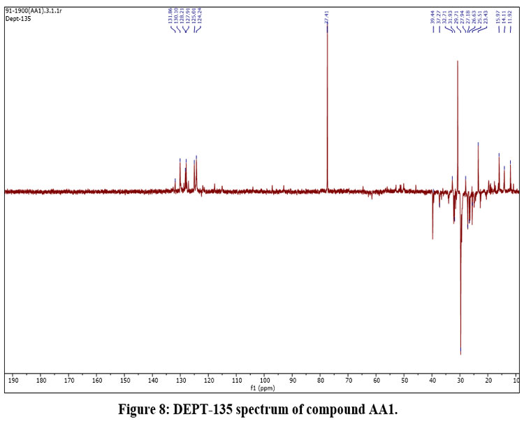 | Figure 8: DEPT-135 spectrum of compound AA1. |
Table 5: 13C NMR and DEPT-135 spectra data of compound AA1
NO. | 13C NMR of AA1 ? (ppm) | DEPT | Remark |
1 | 76.89 | CH | - |
2 | 39.73 | CH2 | - |
3 | 22.70 | CH2 | - |
4 | 29.70 | CH2 | - |
5 | 29.70 | CH2 | - |
6 | 32.70 | CH2 | - |
7 | 135.70 | CH | - |
8 | 128.21 | CH | - |
9 | 128.21 | CH | - |
10 | 128.21 | CH | - |
11 | 32.70 | CH2 | - |
12 | 29.70 | CH2 | - |
13 | 32.70 | CH2 | - |
14 | 22.59 | CH2 | - |
15 | 14.10 | CH3 | - |
1’ | 135.11 | - | Quaternary |
2’ | 127.91 | CH | - |
3’ | 128.21 | CH | - |
4’ | 135.11 | - | Quaternary |
5’ | 128.71 | CH | - |
6’ | 127.91 | CH | - |
7’ | 25.61 | CH3 | - |
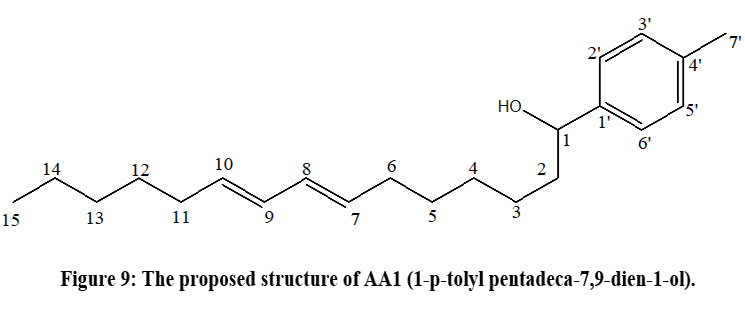 | Figure 9: The proposed structure of AA1 (1-p-tolyl pentadeca-7,9-dien-1-ol). |
Conclusion
In conclusion, n-hexane (4.35 g), chloroform (14.7 g), and methanol (100.7 g) extracts were obtained from Rhus vulgaris. Qualitative phytochemical screening of these crude extracts revealed the presence of alkaloids, glycosides, steroids, anthraquinones, and carbohydrates. Bioactivity assays demonstrated that methanol extracts, containing these bioactive compounds, exhibited significant antibacterial activity, with notable inhibition zones observed. Specifically, the methanol extract showed the highest potency as an antibacterial agent, effectively inhibiting the growth of both Staphylococcus aureus and Salmonella typhi. In contrast, chloroform and n-hexane extracts displayed moderate effectiveness against S. aureus and Escherichia coli, but showed no antibacterial activity against Klebsiella pneumoniae and S. typhi compared to Gentamicin.
Furthermore, the methanol extract was subjected to column chromatography, leading to the isolation and structural elucidation of compound AA1 (1-p-tolyl pentadeca-7,9-dien-1-ol) using chromatographic methods, FTIR, and 1D NMR spectroscopic techniques. These findings highlight the potential of advanced chromatographic techniques such as HPLC and GS-MS for isolating additional compounds from various plant extracts. Additionally, the application of 2D-NMR and MS techniques is recommended for further elucidating the structures of novel compounds isolated from Rhus vulgaris. Future research should include comprehensive bioassay tests on crude extract fractions and isolated compounds to fully evaluate their antibacterial efficacy and potential therapeutic applications.
Acknowledgment
The authors express their sincere appreciation to Abreham Tesfaye Besha for the assistance provided in preparing this manuscript. The authors are grateful for the workshop facilities provided by the University of Gondar (Ethiopia) and Jigjiga University (Ethiopia). I would like to thank the Department of Chemistry at Addis Ababa University and the Faculty of Chemical and Food Science at Bahir Dar University for running the NMR and FTIR spectra analysis of my samples respectively and also the Institute of Biotechnology, the University of Gondar for their contribution in antibacterial analysis.
Funding Sources
The author(s) received no financial support for the research, authorship, and/or publication of this article.
Conflict of Interest
The author(s) do not have any conflict of interest.
Data Availability Statement
This statement does not apply to this article.
Ethics Statement
This research did not involve human participants, animal subjects, or any material that requires ethical approval.
Informed Consent Statement
This study did not involve human participants, and therefore, informed consent was not required.
Authors’ contributions
AA - Study design, Literature search, data collection, data analysis, data interpretation, writing manuscript, GG - Research supervision, Study design, editing manuscript, DS – Research supervision, editing manuscript. All authors read and approved the final manuscript.
References
- Nigussie G, Ashenef S. Isolation, Characterization, Structural Elucidation and Anti-Bacterial Activities of Roots Extracts of Cucumis Ficifolius. Res Sq. Published online 2020:1-31. doi:10.21203/rs.2.22296/v1
CrossRef - Nidal Jaradat, Fatima Hussen, Ali A Al. Preliminary phytochemical screening, quantitative estimation of total flavonoids, total phenols and antioxidant activity of Ephedra alata decne. J Mater Environ Sci. 2015;6(6):1771-1778.
- Mazid M, Khan TA, Mohammad F. Medicinal Plants of Rural India: A Review of Use by Indian Folks. Indo Glob J Pharm Sci. 2012;02(03):286-304. doi:10.35652/igjps.2012.35
CrossRef - Grynkiewicz G, Gadzikowska M. Tropane alkaloids as medicinally useful natural products and their synthetic derivatives as new drugs. Pharmacol Reports. 2008;60(4):439-463.
- Lye KA, Bukenya-Ziraba RR, Tabuti JRS, Waako PJ. Botanical-Medicinal Dictionary for East Africa. Makerere Herb Handb. 2008;(2):1-423.
- Rayne S, Mazza G. Biological activities of extracts from sumac (Rhus spp.): A review. Plant Foods Hum Nutr. 2007;62(4):165-175. doi:10.1007/s11130-007-0058-4
CrossRef - Njoroge PW, Opiyo SA. Antimicrobial Activity of Root Bark Extracts of Rhus natalensis and Rhus ruspoli. Basic Sci Med. 2019;8(2):23-28. doi:10.5923/j.medicine.20190802.01
- Alam P, Parvez MK, Arbab AH, Siddiqui NA, Al-Dosary MS, Al-Rehaily AJ, et al. Inter-species comparative antioxidant assay and HPTLC analysis of sakuranetin in the chloroform and ethanol extracts of aerial parts of Rhus retinorrhoea and Rhus tripartita. Pharm Biol. 2017;55(1):1450-1457. doi:10.1080/13880209.2017.1304428
CrossRef - Gebeyehu G, Asfaw Z, Enyew A, Raja N. Ethnobotanical study of traditional medicinal plants and their conservation status in Mecha Woreda. Int J Pharm Heal Care Res. 2014;02(03):137-153.
- Seble WY, Zemede A, Ensermu K. Ethnobotanical study of medicinal plants used by local people in Menz Gera Midir District, North Shewa Zone, Amhara Regional State, Ethiopia. J Med Plants Res. 2018;12(21):296-314. doi:10.5897/jmpr2018.6616
CrossRef - Mohamed, Khedr FG, Mohammed EI. Phenolic Compounds , Antioxidant and Antibacterial Activities of Rhus flexicaulis Baker. Jordan J Biol Sci. 2019;12(1):17-21.
- Ramathal DC, Ngassapa OD. Medicinal plants used by Rwandese traditional healers in refugee camps in Tanzania. Pharm Biol. 2001;39(2):132-137. doi:10.1076/phbi.39.2.132.6251
CrossRef - Tabuti JRS, Lye KA, Dhillion SS. Traditional herbal drugs of Bulamogi, Uganda: Plants, use and administration. J Ethnopharmacol. 2003;88(1):19-44. doi:10.1016/S0378-8741(03)00161-2
CrossRef - Okullo JBL, Omujal F, Bigirimana C, Isubikalu P, Malinga M, Bizuru E, et al. Ethno-Medicinal Uses of Selected Indigenous Fruit Trees from the Lake Victoria Basin Districts in Uganda. J Med Stud. 2014;2(1):78-88.
- Odongo CO, Musisi NL, Waako P, Obua C. Chewing-stick practices using plants with anti-streptococcal activity in a Ugandan rural community. Front Pharmacol. 2011;2(13):1-5. doi:10.3389/fphar.2011.00013
CrossRef - Ochwangi DO, Kimwele CN, Oduma JA, Gathumbi PK, Mbaria JM, Kiama SG. Medicinal plants used in treatment and management of cancer in Kakamega County, Kenya. J Ethnopharmacol. 2014;151(3):1040-1055. doi:10.1016/j.jep.2013.11.051
CrossRef - Mutuku A, Mwamburi L, Keter L, Ondicho J, Korir R, Kuria J, et al. Evaluation of the antimicrobial activity and safety of Rhus vulgaris (Anacardiaceae) extracts. BMC Complement Med Ther. 2020;20(1):1-12. doi:10.1186/s12906-020-03063-7
CrossRef - Abegaz BM. Novel phenyl anthraquinones, isoflurano naphthoquinones, homoisoflavonoids, and biflavonoids from African plants in the genera Bulbine, Scilla, Ledebouria, and Rhus. Phytochem Rev. 2002;1(3):299-310. doi:10.1023/A:1026066626537
CrossRef - Mdee LK, Yeboah SO, Abegaz BM. Rhus chalcones II-VI, five new bichalcones from the root bark of Rhus pyroides. J Nat Prod. 2003;66(5):599-604. doi:10.1021/np020138q
- Ruffo CK, Birnie A, Tenganäs B. Edible Wild Plants of Tanzania.; 2002.
- Moffett RO. Name changes in the Old World Rhus and recognition of Searsia (Anacardiaceae). Bothalia. 2007;37(2):165-175. doi:10.4102/abc.v37i2.311
CrossRef - Odongo E, Mungai N, Mutai P, Karumi E, Mwangi J, Okalebo F, et al. Antioxidant and anti-inflammatory activities of selected medicinal plants from western Kenya. African J Pharmacol Ther. 2017;6(4):178-182.
CrossRef - Arsule CS. Sable KV. Preliminary Phytochemical Analysis of Euphorbia hirta Linn . Leaves. Int J Life Sci. 2017;5(4):746-748.
- Nataraj ND, B R. PRELIMINARY PHYTOCHEMICAL SCREENING OF SOLANUM TRILOBATUM (L.) YOUNG LEAVES. Int Res J Pharm. 2014;5(2):80-82. doi:10.7897/2230-8407.050216
CrossRef - Vimalkumar CS, Vilash, V. &, Krishnakumar NM. Comparative Preliminary Phytochemical Analysis of Ethanolic Extracts of Leaves of Olea Dioica Roxb, Infected with The Rust Fungus Zahouania oleo Cummins and Non Infected Plants. J Pharmacogn Phytochem. 2014;3(4):69-72.
- Ayoola G, Coker H, Adesegun S, Adepoju-Bello A, Obaweya K, Ezennia E, et al. Phytochemical Screening and Antioxidant Activities of Some Selected Medicinal Plants Used for Malaria Therapy in Southwestern Nigeria. Trop J Pharm Res. 2008;7(3). doi:10.4314/tjpr.v7i3.14686
CrossRef - Savithramma N, Rao ML, Suhrulatha D. Screening of Selected Medicinal Plants for Secondary Metabolites. Middle-East J Sci Res. 2011;8(3):119.
- Khan SA. Antibacterial activity of Rhus succedanea Var. Himalaica. Pure Appl Biol. 2017;6(2):718-724. doi:10.19045/bspab.2017.60076
CrossRef - Khameneh B, Iranshahy M, Soheili V, Bazzaz BSF. Review on plant antimicrobials: a mechanistic viewpoint. Antimicrob Resist Infect Control. 2019;8(1-28). doi:10.1186/s12906-020-03063-7
CrossRef - Gonelimali FD, Lin J, Miao W, Xuan J, Charles F, Chen M, et al. Antimicrobial properties and mechanism of action of some plant extracts against food pathogens and spoilage microorganisms. Front Microbiol. 2018;9:1-9. doi:10.3389/fmicb.2018.01639
CrossRef - Islam T, Islam MN, Zzaman W, Billah MM. Study of Antimicrobial, Antioxidant and Cytotoxicity Properties of Selected Plant Extracts for Food Preservative Applications. Int J Food Stud. 2021;10(February):SI95-SI111. doi:10.7455/ijfs/10.SI.2021.a8
CrossRef

This work is licensed under a Creative Commons Attribution 4.0 International License.